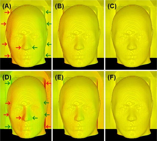
Figure 3.

Volumetric views of two identical CT phantom images with simulated spatial shifts. Top row (A to C): translational shifts (Xt) of 0.5, 0.2 and 0.0 voxels () were applied to the aligned image (C); Bottom row (D to F): rotational shifts (Xr) of 0.5°, 0.2° and 0.0° were applied to the aligned image (F). The color homogeneity on the “skin” landmark improves as the alignment is improved. The translational (lateral) shifts appear mostly on the left and right sides of the image volume: the larger the surface grayscale gradient and the larger the surface oblique angle (between the ray and surface normal), the larger the visual color inhomogeneity would be. The rotational (around the superior‐inferior axis through the center of the image volume) shift causes non‐uniform displacements in the directions perpendicular to the rotational axis: the larger the distance of the viewing voxel to the rotational axis, the bigger the rotational displacement and so the more dramatic color inhomogeneity.
