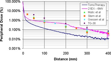
Figure 4. Comparison of peripheral dose distributions measured on a helical tomotherapy system and various conventional 6‐MV beams. The tomotherapy and 21‐EX measurements were taken with a field size. Mutic et al. (9) data measured at with the MLC set to . Stern (10) data measured at a depth of 5 cm with the MLC set to . Giessen (11) averaged measurements for 4‐ to 25‐MV linear accelerators with a field size. TG‐36 values were measured at for a field size of .