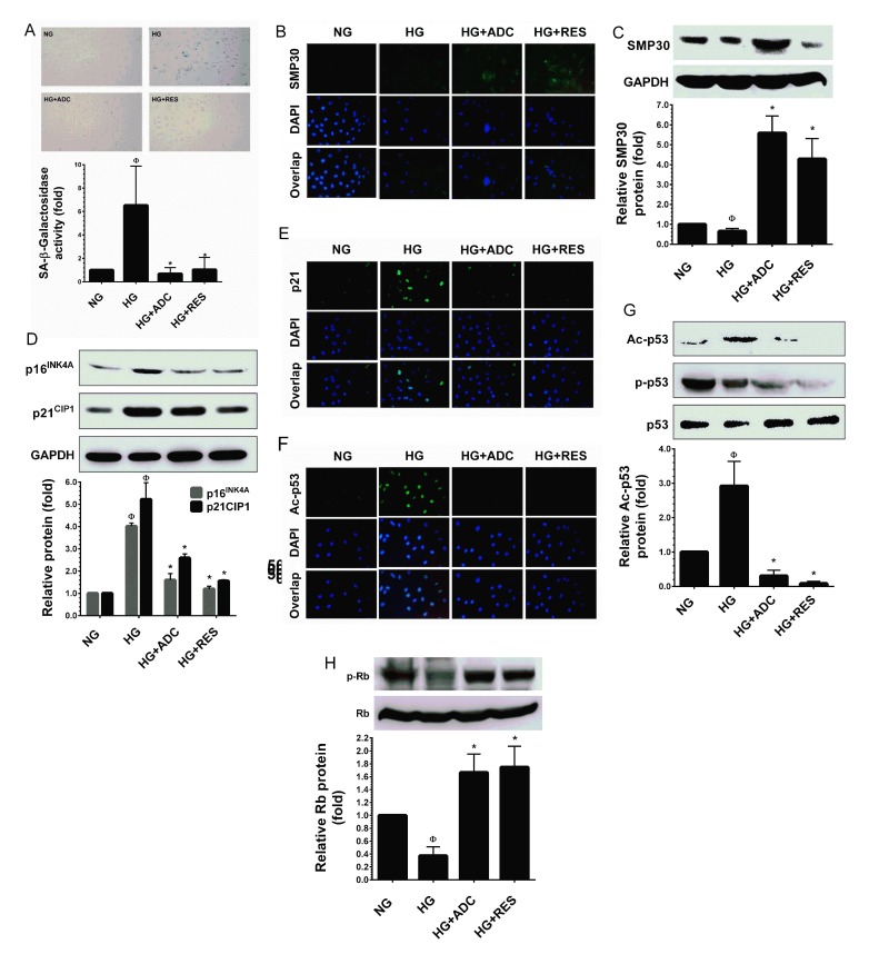
Figure 3. ADC prevents HG-induced senescence in HUVECs.
To determine the effect of ADC on HG-induced senescence, HUVECs were incubated with HG in the presence or absence of ADC or RES for 72 h. A. Cellular senescence was determined by SA-β-gal assay. The top panel shows representative figures and the lower panel shows quantitative analysis of SA-β-gal positive cells per microscopic field. B.,E.,F. The protein expression of SMP30, p21CIP1 and acetylated p53 was measured by immunofluroscence using specific primary antibodies and FITC-conjugated secondary antibody (green). The cellular localization of SMP30, p21CIP1 and acetylated p53 was photographed using a fluorescence microscope. DAPI was used to stain the nucleus. C. SMP30 protein expression level was determined by western blot analysis and the relative SMP30 expression was normalized with GAPDH. D. Senescence-associated protein p21CIP1 and p16INK4A levels were determined by western blotting and the relative protein levels were normalized with GAPDH. G.,H. The protein levels of acetylated p53, phosphorylated p53 and phosphorylated Rb levels were measured by western blot analysis. The relative protein levels of ac-p53 and p-Rb were normalized with total p53 and total Rb levels, respectively. Values represent the mean ± SD of three independent experiments. Statistical significance was set at ФP < 0.05 compared to NG vs. HG and *P < 0.05 compared to HG vs. samples.

