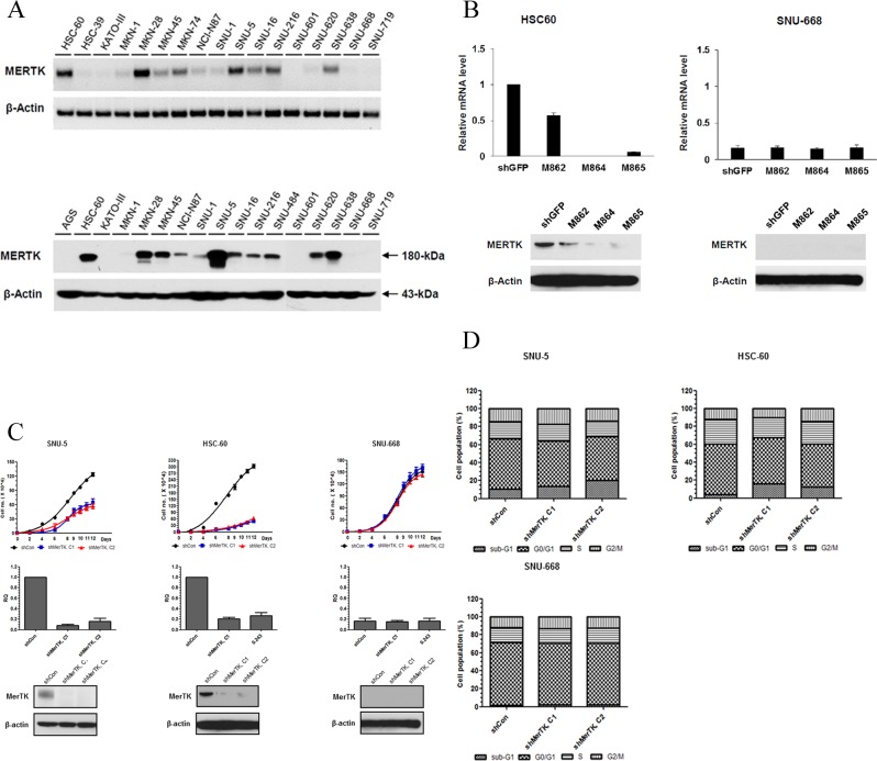
Figure 1. A. Screening for MerTK positive GC cell lines.
(Upper, RT PCR; lower, western blot) B. In HSC-60 (MerTK-overexpressing) cells, shRNA (M864, M865)-mediated knockdown resulted in decreased expression of MerTK mRNA and protein. In SNU-668 cells (MerTK negative), neither the level of mRNA nor protein changed after lentiviral infection. C. The proliferation of two MerTK-positive cell lines (SNU-5 and HSC-60) was significantly inhibited by MerTK targeting shRNA, whereas proliferation of SNU-688 was not affected D. Transfection with MerTK-specific shRNA significantly increased the apoptotic fraction of SNU-5 and HSC-60 cells, compared to a control (M862) clone (16.3%, 12.5%, and 4.1% in cells transfected with M864, M865, and M862, respectively). The apoptotic fraction of SNU-668 cells was unchanged.

