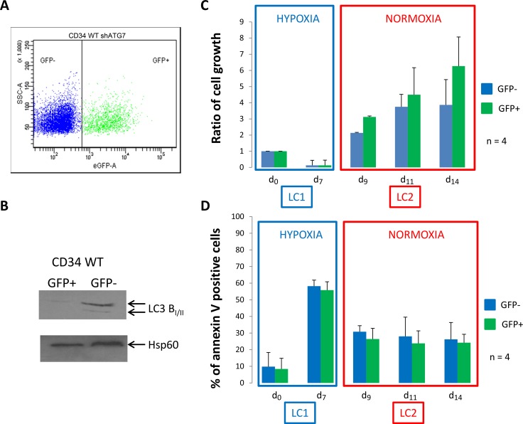
Figure 3. Normal CD34+ progenitor cells proliferate and show low apoptosis back to atmospheric O2 concentration.
(A) Normal CD34+ cells were transduced with a shRNA against ATG7 and analyzed by flow cytometry to determine the percent of GFP- and GFP+ population. (B) Upon cell sorting, GFP- and GFP+ cells were lyzed and autophagy operating mechanism was check by detecting the conversion of microtubule associated light chain 3B-I in LC3B-II. CD34+ cells were cultured at low O2 concentration (0.1% O2) for 7 days (LC1). Upon 7 days, cells were replaced at atmospheric O2 concentration and grown for seven more days (LC2). (C and D) At indicated time, aliquot were analyzed for cell count by trypan blue exclusion assay and apoptosis by flow cytometry using annexin V-APC labelling on the two population GFP- and GFP+. Results are from at least 4 experiments. Significance between autophagy competent and deficient cells was quantitated using Wilcoxon test.

