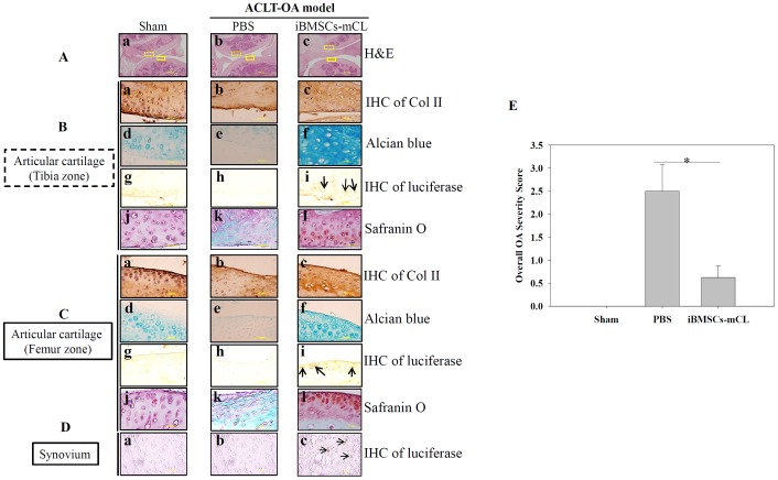
Figure 6. Therapeutic indication of iBMSCs-mCL for OA therapy.
(A) H & E staining of articular cartilage (Aa-c) (100X) in tibia zone ( ) and femur zone (
 ) in sham, PBS and iBMSCs-mCL groups. (B) and (C) Staining of articular cartilage in tibia zone and femur zone respectively. Type II collagen (Col II) (tibia zone: Ba-c; femur zone: Ca-c), Alcian blue staining for proteoglycans (tibia zone: Bd-f; femur zone: Cd-f) and IHC staining of luciferase (tibia zone: Bg-i; femur zone: Cg-i; arrows indicated) (1000X), safranin o staining (tibia zone: Bj-l, femur zone: Cj-l). (D) IHC staining of luciferase (synovium: Da-c). (E) Comparative histologic scores of OA among sham, PBS and iBMSCs-mCL groups. * denotes the significant difference as P<0.05.

