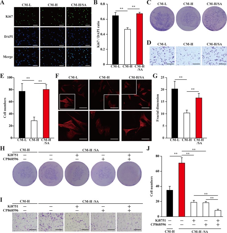
Figure 5. Salidroside promotes smooth muscle cells proliferation and migration potentials via skeletal cells secretome.
(A, B) The proliferation of MOVAS cells cultured with indicated conditioned media, as analyzed by Ki67 staining: (A) representative images (scale bars: 200 μm); and (B) quantification of the ratio of Ki67 positive cells to DAPI positive cells. (C) The number of MOVAS cells cultured with indicated conditioned media, as analyzed by crystal violet. (D, E) The mobility of MOVAS cells cultured with indicated conditioned media was examined using transwell chamber assay: (D) representative images (scale bars: 100 μm); and (E) quantification of migrated cells. (F, G) Morphological changes of F-Actin, as examined by phalloidin staining: (F) representative images (scale bars: 100 μm for upper panels and 50 μm for lower panels), lower panels showed the enlarged images of the cropped part in the upper panels; (G) quantification analysis of fractal dimension. (H) The number of MOVAS cells cultured with CM-H/SA and indicated inhibitors, as analyzed by crystal violet staining. (I, J) The mobility of MOVAS cells cultured with CM-H/SA and indicated inhibitors, as examined by transwell chamber assay: (I) representative images (scale bars: 100 μm); and (J) quantification of migrated cells. All experiments were done under hypoxia. Data shown are representative from three independent experiments. Quantification data were expressed as mean ± S.D. (n = 6). **P < 0.01. CM-L: conditioned medium collected from C2C12 cells treated with PBS and cultured under low glucose condition; CM-H: conditioned medium collected from C2C12 cells treated with PBS and cultured under hyperglycemia; and CM-H/SA: conditioned medium collected from C2C12 cells treated with salidroside and cultured under hyperglycemia; Ki8751: VEGFR inhibitor; CP868596: PDGFR-B inhibitor.

