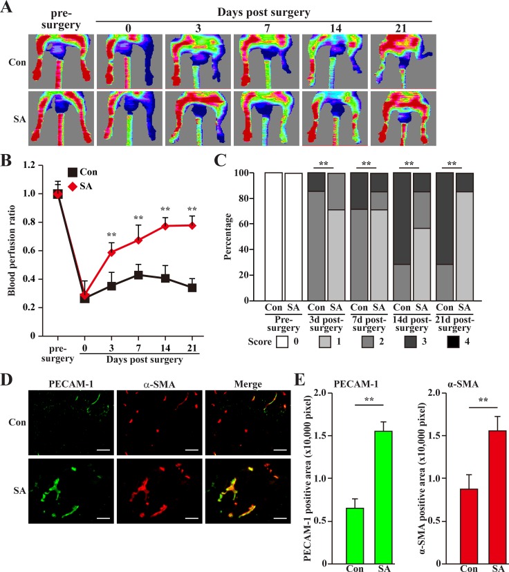
Figure 8. Salidroside promotes blood perfusion recovery by inducing neoangiogenesis in diabetic HLI model mice.
(A) Representative of Laser Doppler Perfusion Images showing blood perfusion in the ischemic and non-ischemic hind limb of diabetic HLI model mice treated with PBS (upper panels) or salidroside (lower panels). (B) The blood perfusion ratio of ischemic hind limb to non-ischemic hind limb. Data were shown as mean ± S.D. (n = 7 per group). (C) Assessment test of the ischemic hind limb morphologies of diabetic HLI model mice treated by salidroside or PBS at indicated times (n = 7 per group, 0 = no difference from the non-ischemic hind limb, 1 = mild discoloration, 2 = moderate discoloration, 3 = severe discoloration, subcutaneous tissue loss, or necrosis, and 4 = amputation). (D, E) Immunohistochemistry against PECAM-1 (green) and α-SMA (red) in the gastrocnemius muscle of the ischemic hind limbs of diabetic HLI model mice treated by either salidroside or PBS at 21 days post-surgery: (D) representative images (scale bars: 100 μm); (E) quantification of PECAM-1 (left panel) and α-SMA positive (right panel) areas. Data shown are representative from three independent experiments. Quantification data were shown as mean ± S.D. (n = 6). **P < 0.01 (control versus salidroside-treated); Con: animals treated with PBS; SA: animals treated with salidroside.

