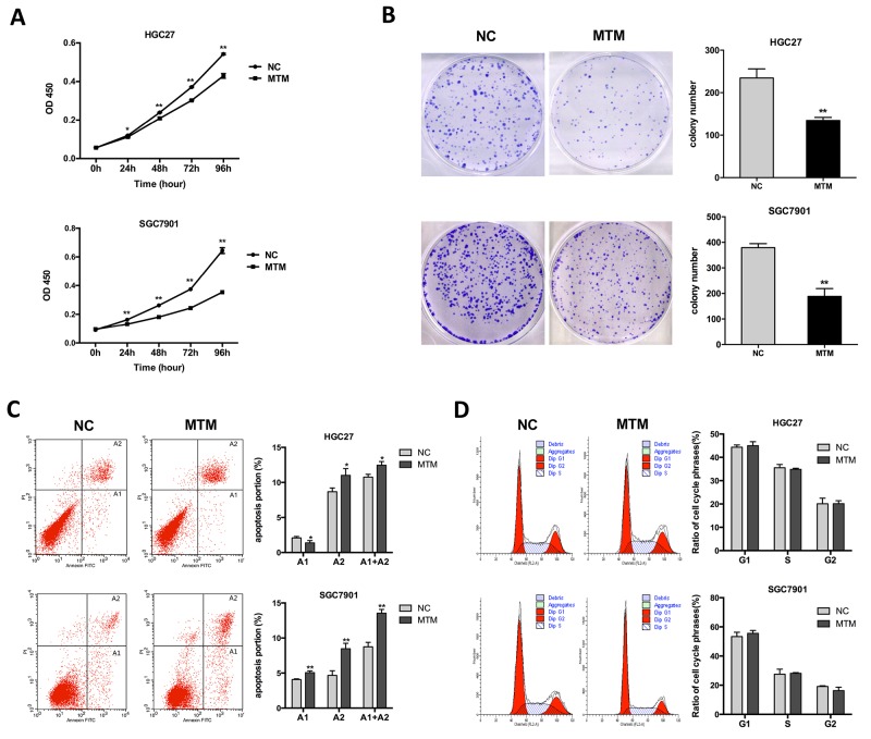
Figure 3. Effects of MTM overexpression on gastric cancer (GC) cell proliferation.
(A) CCK-8 assays showed the proliferation of GC cells was inhibited by MTM overexpression. Data are represented as mean ± SD (n=5, *P<0.05, **P<0.01). (B) Representative pictures of colony formation in MTM-overexpressing GC cells. The histogram showed the average number of the survival clones. Data were presented as the mean ± SD (n=3, **P<0.01). (C) The apoptotic rates of cells were detected by flow cytometry in transfected GC cells (A1, early apoptosis; A2, late apoptosis). Data represented the mean ± SD (n=3, *P<0.05, **P<0.01). (D) Cell cycle distribution was analyzed by flow cytometry in GC cells. Data were presented as the mean ± SD (n=3, all P>0.05).

