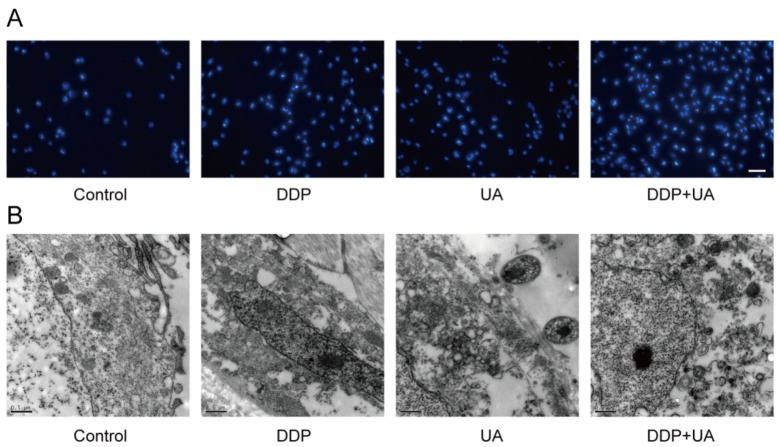
Figure 4. UA promoted DDP-induced SiHa cell morphological changes.
(A) Determination of apoptosis in the SiHa cells 24h post DDP in the presence of UA was analyzed by Hoechst 33258 staining method (×200). (B) The ultrastructural morphological investigation of SiHa cells using TEM 72h after DDP treatment in the presence of UA (0.5μm). DDP: cisplatin; UA: ursolic acid.

