
Figure 7. UA restrained DDP-induced NF-κB p65 activation in SiHa cells.
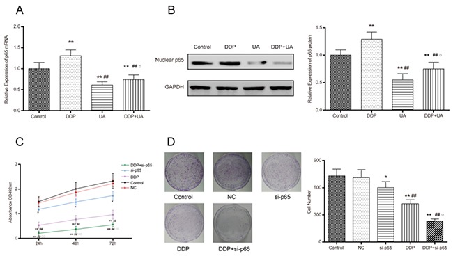
(A) The expression level of the mRNA nuclear NF-κB p65 was measured by RT-PCR. (B) The expression level of the protein nuclear NF-κB p65 was measured by western blot and RT-PCR. Results (mean ± SD) were from six independent experiments. ** P < 0.01 versus the control group; ## P < 0.01 compared with the DDP group; ○P < 0.05 compared with the UA group. (C) Cell proliferation was measured by MTT assay. (D) DDP and si-p65 affected cell colony formation in SiHa cells. Results (mean ± SD) were from six independent experiments. * P < 0.05 versus control group; **P < 0.01 versus the control group; ## P < 0.01 compared with the si-p65 group; ○P < 0.05 compared with the DDP group. NC: cells transfected with empty vector plasmid. si-p65: cells transfected with vector of si-p65 sequences. DDP + si-p65: cells transfected with vector of si-p65 sequences and treated with DDP. DDP: cisplatin; UA: ursolic acid.