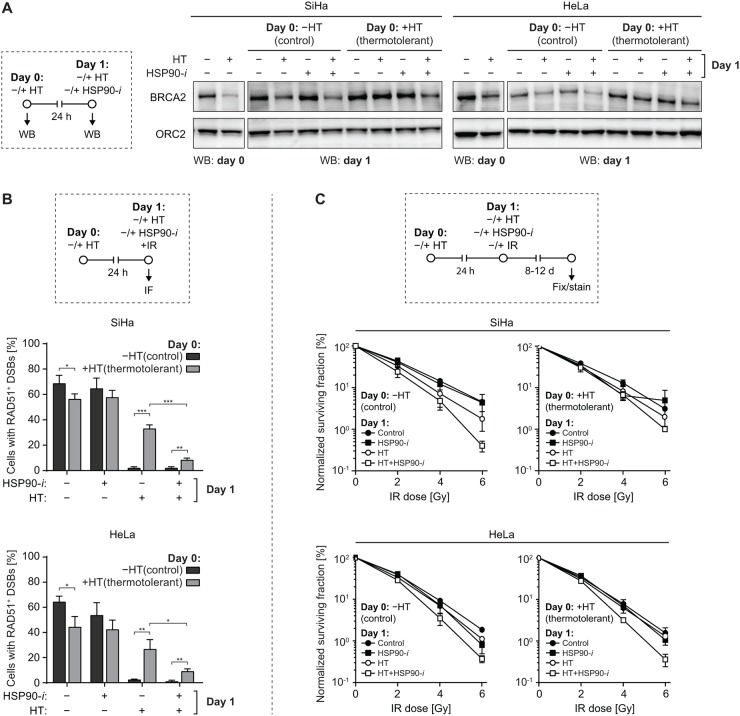
Figure 5. Inhibition of HSP90 reduces thermotolerance.
(A) SiHa or HeLa cells were sham-treated or rendered thermotolerant by incubation for 1 h at 37 or 42°C (HT), respectively. Some of the treated cell samples were immediately lysed and analyzed by Western blotting to confirm the effectiveness of hyperthermia (HT) treatment by analyzing BRCA2 levels (panels marked by ‘WB: day 0’). One day later, the remaining samples were subjected to a second HT treatment, in the absence or presence of 30 nM Ganetespib (HSP90-i), lysed, and BRCA2 levels were determined by Western blotting (panels marked by ‘WB: day 1’). The experimental design is schematically represented in the left-hand panel. ORC2 was used as a loading control. (B) SiHa or HeLa cells were treated as described in (A), except immediately after the second HT treatment cells were irradiated using α-particles, fixed 30 min later and immunostained against γH2AX and RAD51. Graphs represent the average percentage of cells containing α-particle induced tracks of γH2AX foci that were also positive for RAD51. The experimental design is schematically represented in the top panel. (C) Cells were rendered thermotolerant as in (A). 24 h later the cells were trypsinized, counted and seeded into 6-well plates. 4 h after seeding, they were incubated for 1 h at 37 or 42°C, in the absence or presence of HSP90-i, and exposed to the indicated dose of IR. Normalized clonogenic survival was determined 8 (HeLa) or 12 (SiHa) days later. The experimental design is schematically represented in the top panel.

