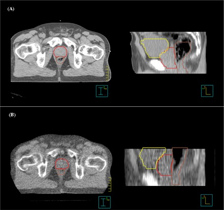
Figure 1.

Comparison of (A) diagnostic computed tomography (CT) and (B) megavoltage CT images taken on the HI‐ART2 system (TomoTherapy, Madison, WI) for axial (left panel) and sagittal (right panel) views. The prostate, rectum, and bladder contours are shown. Note the superior–inferior displacement of the prostate relative to the reference treatment‐planning CT.
