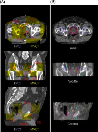
Figure 2.

The image fusion tool on the TomoTherapy (Madison, WI) operation station is used to register the treatment planning kilovoltage computed tomography (kVCT) images with megavoltage computed tomography (MVCT) images. After an automated CT‐to‐CT fusion, the operator can manually adjust the registration (A) by using a “checkerboard” display or (B) by overlaying the treatment planning contours with the MVCT image. Axial, sagittal, and coronal views are shown for both registration techniques. Arrows in the “checkerboard” display point to anatomic landmarks used in manual adjustments for prostate patients. The prostate (red), planning target volume (pink), rectum (brown), bladder (yellow), and femoral heads (blue) are shown in the contour overlay. ; ; ; ; .
