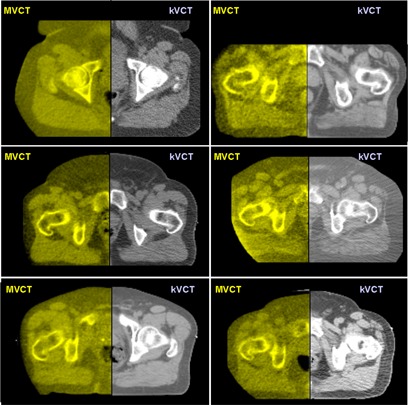
Figure 6.

Six random examples of axial megavoltage computed tomography (MVCT)–to–kilovoltage computed tomography (kVCT) registration for prostate patients using the “checkerboard” image fusion tool on the TomoTherapy (Madison, WI) operator station. The right half of each prostate image is the reference kVCT (grayscale), and the left half is the daily MVCT image (yellowscale). Note that the prostatic–rectal interface is visible in each image.
