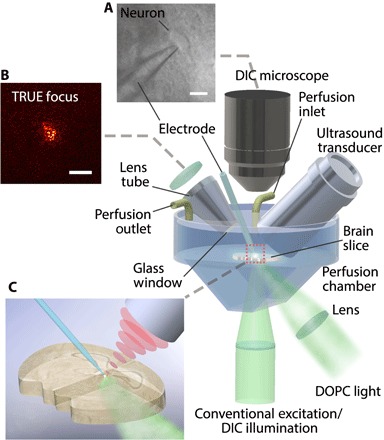
Fig. 1. Custom TRUE focusing and electrophysiological recording system.

The custom TRUE focusing system combined a DOPC system with a patch clamp electrophysiology amplifier and headstage. Acute brain slices were held in a custom perfusion chamber that contained warmed, carbogenated aCSF. The TRUE light beam illuminated the tissue at an oblique 45° angle, and the borosilicate patch pipette electrode was used for neurophysiological measurements. (A) A DIC microscope was included for neuron visualization during patch clamping. (B) The TRUE focusing system allowed light to be sharply focused through the brain slice. (C) A close-up image of the TRUE focus on a patched neuron. Scale bars, 20 μm in (A) and 50 μm in (B).
