
Fig. 4. 3D volume reconstruction of 10-μm fluorescent beads suspended in agarose.
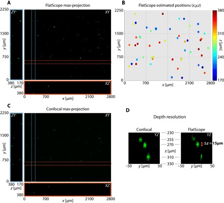
(A) FlatScope reconstruction as a maximum intensity projection along the z axis as well as a ZY slice (blue box) and an XZ slice (red box). (B) Estimated 3D positions of beads from the FlatScope reconstruction. (C) Ground truth data captured by confocal microscope (10× objective). (D) Depth profile of reconstructed beads compared to ground truth confocal images. Empirically, we can see that the axial spread of 10-μm beads is around 15 μm in FlatScope reconstruction. That is, FlatScope’s depth resolution is less than 15 μm. The three beads shown are at depths of 255, 270, and 310 μm from the top surface (filter) of the FlatScope.