
Figure 3. Increased inflammatory cytokines and mediators resulting from deletion of myeloid specific TGFβ signaling.
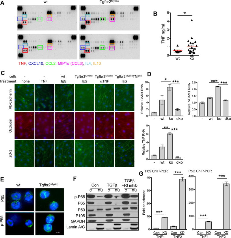
(A) Cytokine antibody array of plasma from one male and one females 6 month-old pre-stroke Tgfbr2Myeko mice, compared one male and one female wt (n=2 for each group). Boxed cytokines are indicated with same color text below. (B) TNF ELISA of plasma from 6–8 month wt and Tgfbr2Myeko mixed male and female mice pre-stroke (n=10 wt and n=20 ko). (C) Tgfbr2Myeko myeloid cells cause interruption in adherens junctions and tight junctions between co-cultured C57BL/6 mouse brain microvascular endothelial cells. Co-culture with peripheral blood myeloid cells (cells) from wt, Tgfbr2Myeko or Tgfbr2MyekoTNF−/− (ko) mice. Some samples were treated with TNF, aTNF neutralizing antibody or IgG isotype antibody. Notice disruption in the endothelial junctions with TNF treatment and by Tgfbr2Myeko myeloid cells. (D) C57BL/6 brain microvascular endothelial cells have elevated RNA for markers of endothelial damage (top panels) and TNF (lower panel) following co-culture with Tgfbr2Myeko (ko) myeloid cells that can be reduced when myeloid cells lack TNF also (dko, Tgfbr2MyekoTNF−/−). (E) Increased NFkB transcription factor subunit P65 (encoded by Rela) and active, phosphorylated P65 in myeloid cells from Tgfbr2Myeko mice. P65 and p-P65 (green), nuclei (blue, Dapi). Shown are representative pictures. (F) Increased nuclear NFkB subunits P65 and P50 (encoded by Nfkb1) and active p-P65 upon blockade of TGFb signaling using a TbR1 (R1) inhibitor. Western blots of nuclear (nu) and cytoplasmic (c) fractions from RAW264.7 cells. (G) Increased binding of NFkB to the TNF promoter after knock-down (KD) of Tgfbr2. Fold enrichment of TNF from P65 Chromatin immunoprecipitation (ChIP) of RAW264.7 cells. TNF1 or TNF2 indicates two different regions in the TNF promoter (see Online Figure 3C).