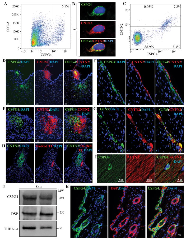
Figure 1. Detection of expression of CSPG4 in the mouse heart and skin.
A. Flow cytometry plot showing CSPG4pos isolated by FACS from the mouse non-myocyte cardiac cell fraction. CSPG4pos cells represented 5.6±3.3% (N=6) of the non-myocyte cells in the mouse heart. Corresponding controls are shown in Online Figure 1. B. Co-immunostaining of cardiac cells isolated by flow cytometry for the expression of CSPG4 (validation) and cardiac conduction system (CCS) marker contactin 2 (CNTN2). As shown CSPG4 and CNTN2 are co-expressed in the isolated cells. C. Flow cytometry plot showing sorting of cardiac non-myocyte cells against anti CSPG4 and CNTN2 antibodies. As shown, approximately 2/3rd of the cells expressing CSPG4 also expressed CNTN2. D and E. Co-immunofluorescence staining of thin myocardial sections for CSPG4 and CNTN2 showing expression of the CSPG4 protein in in the AV nodal area, identified by the expression of the CCS marker CNTN2. Lower (D) and higher (E) magnifications of the AV nodal area are shown. F and G panels illustrate co-expression of CSPG4 and CNTN2 (Panel F) or GJN5 (connexin 40) and CNTN2 (Panel G) in the CCS, likely representing one of the bundle branches. H. Thin myocardial sections from the Cspg4-DsRedT.1 reporter mouse showing expression of Ds-Red.T.1 protein, a surrogate for CSPG4, in the AV nodal area, which is also identified by the expression of CNTN2. I. Isolated CSPG4pos cells within the myocardium residing between myofibrillar bundles, likely representing a pericytes or a neuroglial type II cells. The panel also shows absence of expression of CSPG4 in cardiac myocytes. J. Immunoblot showing expression of CSPG4 and DSP proteins in the mouse skin tissue along with a corresponding control for the loading condition. K. Thin skin sections showing co-expression and co-localization of CSPG4 and DSP in the epidermis and around hair follicles.

