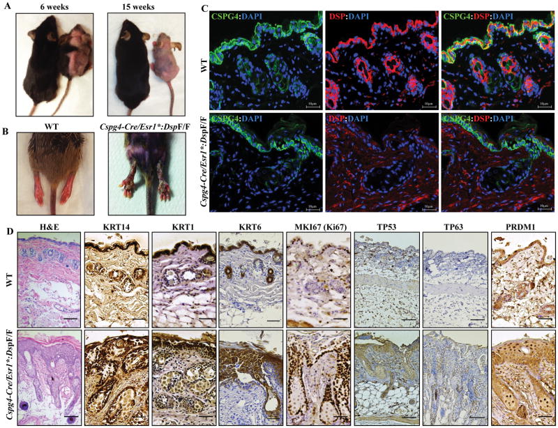
Figure 6. Alopecia totalis, palmoplantar keratosis, increased proliferation and terminal differentiation of epidermal keratinocytes upon deletion of Dsp in the CSPG4pos cells.
A. Representative images of WT and Cspg4-Cre/Esr1*:DspF/F mice illustrating progressive hair loss (alopecia) in the Cspg4-Cre/Esr1*:DspF/F mouse at 6 (left panel) and 15 weeks of age (3 and 12 weeks post tamoxifen-induced deletion of Dsp). B. Representative image showing the presence of severe keratosis on the hind legs of Cspg4-Cre/Esr1*:DspF/F mice. C. Thin skin sections obtained from WT and Cspg4-Cre/Esr1*:DspF/F mice stained for the expression of CSPG4 and DSP proteins. CSPG4 was expressed in the epidermal layer and around hair follicles in both genotypes. Likewise, DSP was expressed and localized to epidermal keratinocytes and hair follicles in the WT but was absent in the Cspg4-Cre/Esr1*:DspF/F mice. D. Immunohistochemical panels of skin tissue from WT and Cspg4-Cre/Esr1*:DspF/F mice showing expression of selected markers for proliferation and differentiation. H&E stained section shows thickened epidermis and perturbed epidermal compaction and sheet formation. KRT14, TP63 and MKI67 (Ki67) stained panels show a hyperproliferative epidermal basal layer, while KRT1, KRT6, TP53 and PRDM1 (BLIMP1) stained panels show impaired terminal differentiation of epidermal keratinocytes.

