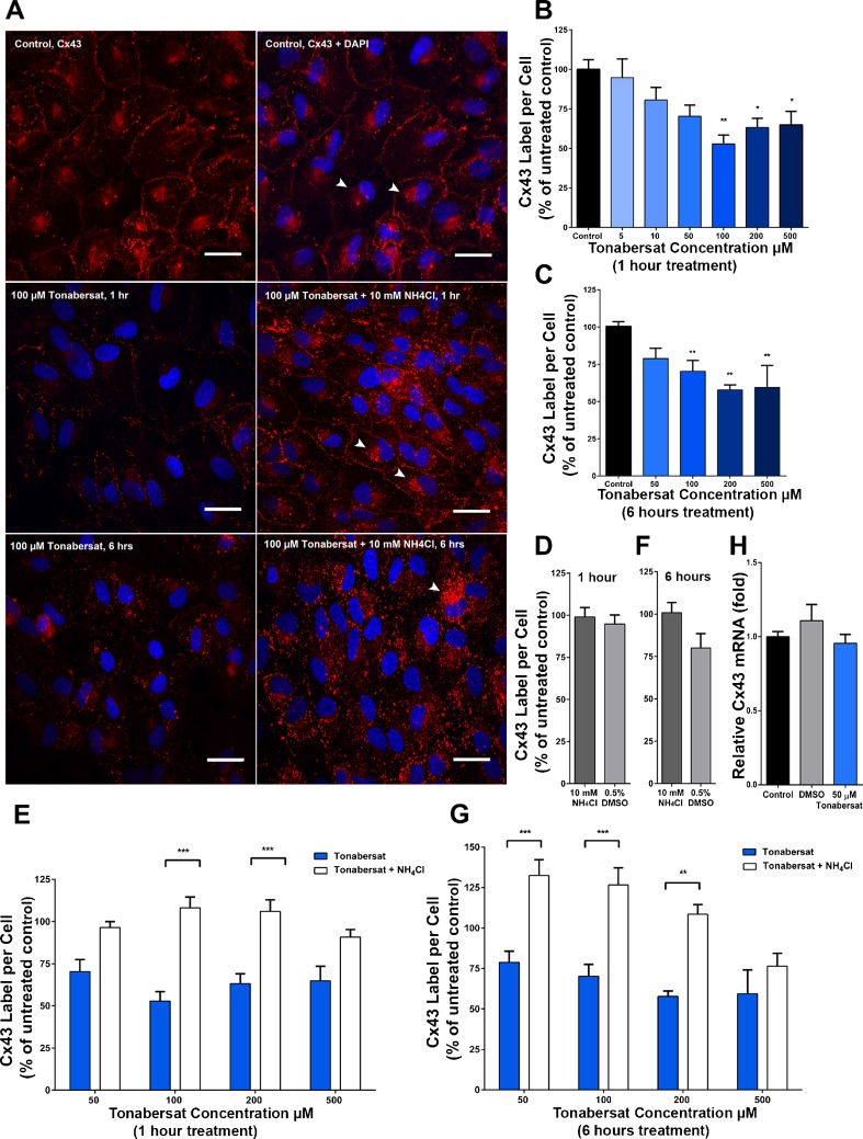
Fig. 3.
Tonabersat internalizes and degrades connexin43 gap junction plaques via the lysosomal pathway in ARPE-19 cells. (A) Immunolabeling for connexin43 (Cx43; red) and 4',6-diamidino-2-phenylindole (DAPI)-labeled nuclei (blue) in untreated control cells (upper panel), after 100 μM tonabersat for 1 h (middle left), 100 μM tonabersat + NH4Cl for 1 h in serum-supplemented media (middle right), 100 μM tonabersat for 6 h (lower left), or 100 μM tonabersat + NH4Cl for 6 h (lower right). White arrows indicate perinuclear labeling of connexin43; scale bars = 30 μm. Quantification of the total area of connexin43 plaques per cell following treatment with tonabersat (5–500 μM) for 1 h is seen in (B), and tonabersat (50–500 μM) for 6 h in (C). (B, C) Normalized to untreated control and expressed as a percentage. Quantification of the total area of connexin43 plaques per cell following treatment with tonabersat (50–500 μM) with NH4Cl for 1 h is seen in (E) and for 6 h in (G) with both figures normaliszed to untreated control and expressed as a percentage. NH4Cl prevents junctional degradation, indicating that it would otherwise occur via the lysosomal pathway. No significant difference in connexin43 labeling was present between untreated control and 10 mM NH4Cl or vehicle control [0.5% dimethyl sulfoxide (DMSO)] alone for (D) 1 h or (E) 6 h. Confluent monolayers of ARPE-19 cells were incubated with DMSO (vehicle control), or with 50 μM tonabersat for 1 h and relative connexin43 mRNA levels were then assessed. (H) Tonabersat has no effect on connexin43 transcription. Values represent mean ± SEM. One-way analysis of variance followed by Tukey’s multiple comparisons test. *p < 0.05; **p < 0.01 ***p < 0.001; n = 3 wells per treatment for each of 2 independent experiments

