Figure 3.

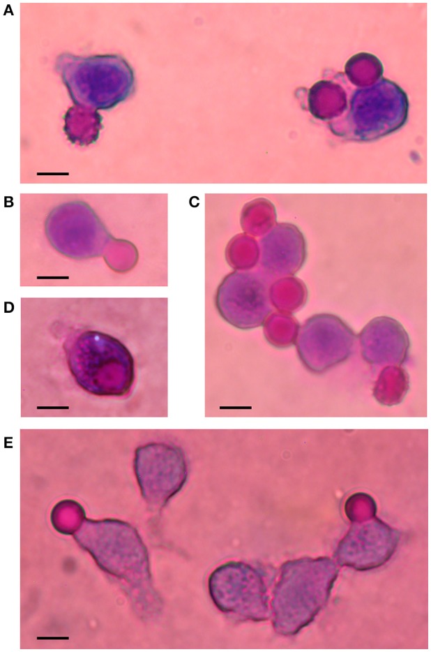
Phagocytosis of Ca2+-loaded RBCs. Illustrative images of Wright-stained cells are shown in this composite figure. Ca2+-loaded RBCs (7 μM) were incubated for 2 h in RPMI medium, in the absence (A) and presence of AS (B), affinity-purified IgG (C), IgG-depleted serum (D) and inactivated IgG-depleted serum (E). Notice late stages of echinocytes: crenated and smooth spheres, undergoing phagocytosis. Bars represent 10 μm.
