Figure 5.
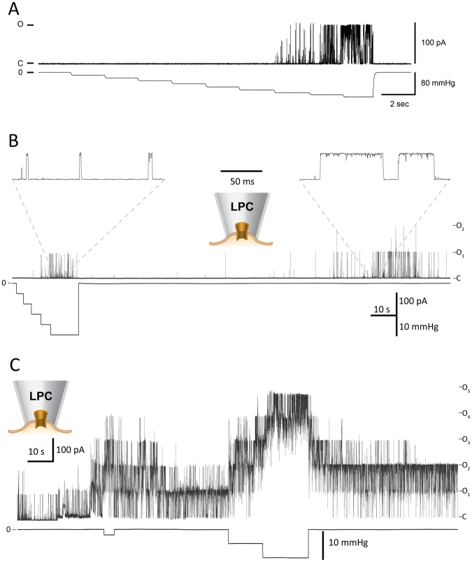
Patch-clamp recording from purified MscL mutant V120C reconstituted into liposomes. (A) The MscL-V120C mutant current recorded upon the application of 10 mmHg suction steps onto the patch area. The channel currents (top) and the negative pressure applied to the inside-out azolectin liposome patch through the patch pipette (bottom) are shown. (B) Activation of the mutant channel by application of 3 μM LPC in the patch pipette, which is much less than 25 mol% used in EPR experiments (Fig. 4). (Note that it took ∼2 min for LPC to diffuse inside the pipette to reach and incorporate in the monounsaturated (18:1) POPC liposome patch and activate the channel.) The channel activity is characterized by brief channel openings. Expanded views show the openings of the MscL-V120C mutant in the presence (left) and absence of suction (right) after LPC incorporation into the liposome patch. (C) The activation of the MscL mutant channel in the presence of 5 μM LPC in the patch pipette before, during and after application of suction to the pipette. Note longer openings of multiple channels when compared to 3 μM LPC recordings. Pipette potential was +30 mV.

