Figure 5.
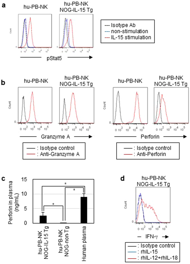
Analysis of functions in in vivo-expanded human PB-NK cells. (a) Phosphorylation of STAT5. Freshly isolated hu-PB-NK cells and NK cells in NOG-IL-15 Tg mice at 6 weeks after transfer were stimulated with recombinant human IL-15 (2 ng/ml) for 30 min at 37 °C. The cells were analyzed by phosflow. (b) Production of granzyme A and perforin molecules. The cells were cultured in the presence of 3 μg/ml breferdin A for 20 h at 37 °C, and subsequently fixed, permeabilized, and stained with indicated antibodies specific for granzyme A or perforin. The fresh PB-NK cells and in vivo-expanded NK cells in NOG-IL-15 Tg mice were derived from different donors. (c) Quantitation of human perforin in plasma. Plasma was prepared from the PB of NOG-non-Tg (n = 12) or NOG-IL-15 Tg (n = 15) mice at 6 weeks after transfer of hu-PB-NK cells. The plasma from normal human donors (n = 5) was used as a control. Perforin levels were determined by ELISA.The p-value was obtained using Student’s t-test (*p < 0.01). (d) IFN-γ production of in vivo-expanded hu-PB-NK cells. Hu-PB-NK cells prepared from NOG-IL-15 Tg mice at 6 weeks post-transfer were stimulated with recombinant human IL-15 or a combination of human IL-12 and IL-18 in the presence of breferdin A. After culture for 20 h at 37 °C, the cells were intracellularly stained with anti-human IFN-γ. For FACS plots, a representative result from triplicate samples for each staining group is shown. The experiments were repeated twice using different mice and human donors.

