Fig. 1.
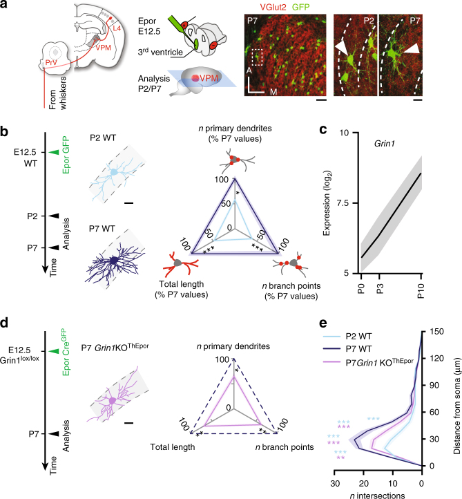
NMDAR activation controls the dendritic maturation of VPM neurons during postnatal development. a Left: somatosensory thalamocortical neurons of the ventroposterior medial nucleus (VPM) of the thalamus respond to whisker inputs and project to the primary somatosensory cortex. Center: experimental time course and schematic representation of the labeling technique. Right: GFP+ neurons are visible in horizontal sections of the VPM in the low magnification photomicrograph and VGlut2 labeling allows delineation of the barreloids. Scale bar: 100 μm. High magnification of two VPM neurons at P2 and P7 (arrowheads) with white dotted lines delineating barreloids. Scale bar: 20 μm. b Dendritic complexity of VPM neurons increases during development (P2 WT n = 11 from 2 mice, P7 WT n = 10 from 2 mice). Three-axis representation of primary dendrites, number of branch points and total dendritic length. Values are expressed as a percentage of WT P7 VPM neurons values in this and subsequent panels. See Supplementary Fig. 2 for related data. Scale bar: 20 μm. c Grin1 expression increases during development. d Dendritic maturation is impaired in Grin1 loss-of-function neurons (P7 Grin1KOThEpor n = 16 from 3 mice). Scale bar: 20 μm. e Sholl analysis of dendritic complexity. PrV, principal trigeminal nucleus; WT, wild-type. One-way ANOVA with Tukey's post-hoc test for all statistical tests relating to dendritic complexity, except for Sholl analyses for which a two-way ANOVA with Tukey post-hoc test was used. *P < 0.05, **P < 0.01, ***P < 0.001; NS, not significant

