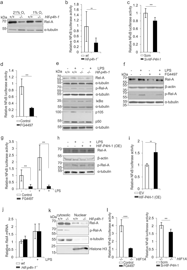
Figure 3.
Lack of HIF-P4H-1 suppresses NF-κB activity. (a) Western blot analysis of Rel-A in wt and Hif-p4h-1 −/− MEFs subjected to 21% or 1% O2 for 24 h. (b–d) Analysis of NF-κB luciferase reporter activity in wt and Hif-p4h-1 −/− MEFs (b), in Hif-p4h-1 and scrambled (Scrm) siRNA transfected HEK293 cells (c) and in MDA-MB-231 cells treated with 50 µM FG4497 for 6 h (d). Cells were harvested 48 h after the NF-κB luciferase reporter plasmid transfection. siRNA transfection was performed 24 h before the NF-κB luciferase reporter plasmid transfection. FG4497 was added 6 h before the cell harvest. (e) Western blot analysis of Rel-A, p-Rel-A, IκBα, p105 and p50 in wt and Hif-p4h-1 −/− MEFs treated with or without 200 ng/ml LPS for 12 h. (f,g) Analysis of Rel-A and p-Rel-A by Western blotting (f) and NF-κB luciferase reporter activity (g) in wt MEFs treated with or without 200 ng/ml LPS and 50 µM FG4497 for 6 h. (h,i) Analysis of Rel-A and p-Rel-A by Western blotting (h) and NF-κB luciferase reporter activity (i) in Hif-p4h-1 −/− MEFs transfected with empty vector (EV) or a vector encoding V5-tagged human recombinant HIF-P4H-1 (OE) and treated with or without 200 ng/ml LPS for 12 h. (j) qPCR analysis of Rel-A mRNA in Hif-p4h-1 −/− and wt MEFs treated with 200 ng/ml of LPS for 12 h. (k) Western blot analysis of Rel-A and p-Rel-A in cytosolic and nuclear fractions of Hif-p4h-1 −/− and wt MEFs. (l) NF-κB luciferase reporter activity in HIF1A −/− HCT116 cells treated with FG4497 or transfected with HIF-P4H-1 siRNA. Data are presented as representative Western blots and as mean ± s.d., n = 3–4 individual MEF isolates or experiments. *P < 0.05, **P < 0.01 and ***P < 0.001, two-tailed Student’s t-test. Unprocessed original scans of blots are shown in Supplementary Fig. 5.

