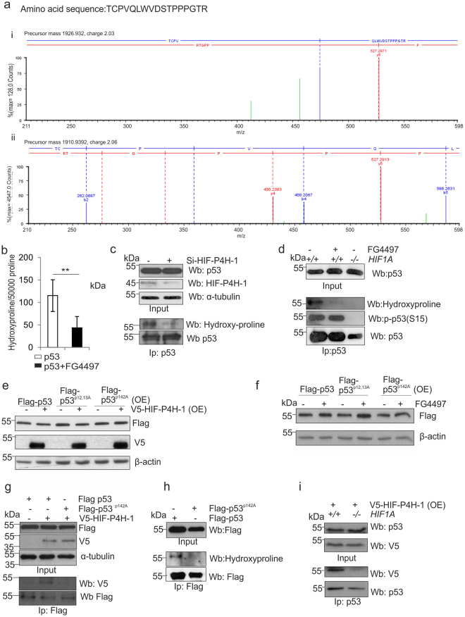
Figure 7.
p53 is hydroxylated at Pro142. (a) Flag-tagged p53 was immunoprecipitated from HCT-116 cells treated with 10 μg/ml cisplatin and 10 μM MG132 for 6 h, separated by SDS-PAGE, trypsinized and analyzed by LC-MS. Mass signals extracted from the high energy trace of the MSe measurement assigned to the peptide TCPVQLWVDSTPPPGTR (140–156) are shown. Low molecular weight area for the oxidized (i) and unmodified (ii) peptide with the b4 ions was used to assign hydroxylation to Pro142. Detailed information on all found fragment ions are provided in Supplementary tables 5 and 6. (b) [2,3,4,5-3H] proline-labeled Flag-tagged p53 was produced in wt HCT-116 cells treated with 10 μg/ml cisplatin and 10 μM MG132 with or without 50 μM FG4497 for 6 h. Flag-p53 was immunoprecipitated and the amount of 4-hydroxy[3H]proline formed was measured by a radiochemical method. The data are given as the amount of 4-hydroxyproline residues/50 000 proline residues. (c) HCT-116 cells were transfected with scrambled or HIF-P4H-1 siRNA and treated with 10 μg/ml cisplatin and 10 μM MG132 for 6 h. Endogenous p53 was immunoprecipitated and analysed by Western blotting with antibodies agains hydroxyproline and p53. (d) Wt and HIF1A −/− HCT-116 cells were treated with 10 μg/ml cisplatin and 10 μM MG132 for 6 h with or without 50 μM FG4497. Endogenous p53 was immunoprecipitated and analysed by Western blotting with antibodies against hydroxyproline, P(Ser15)-p53 and p53. (e,f) Flag-tagged wt, Pro12Ala/Pro13Ala and Pro142Ala p53 were overexpressed (OE) in HCT-116 cells with or without V5-tagged HIF-P4H-1 (e) and with or without 50 μM FG4497 for 6 h (f) and the amount of p53 and HIF-P4H-1 was analysed by Western blotting with anti-Flag and anti-V5 antibodies, respectively. (g) Flag-tagged wt and Pro142Ala p53 and V5-tagged HIF-P4H-1 were overexpressed in HEK293 cells and treated with 10 μM MG132 for 6 h. Flag-tagged p53 proteins were immunoprecipitated and co-immunoprecipitation of HIF-P4H-1 was analysed by Western blotting with an anti-V5 antibody. (h) Flag-tagged wt and Pro142Ala p53 were overexpressed in HCT-116 cells and treated with 10 μg/ml cisplatin and 10 μM MG132 for 6 h. Flag-p53 was immunoprecipitated and hydroxylation was analysed by Western blotting with an anti-hydroxyproline antibody. (i) V5-tagged HIF-P4H-1 was overexpressed (OE) in wt and HIF1A −/− HCT-116 cells treated with 10 μM MG132 for 6 h. Endogenous p53 was immunoprecipitated and co-immunoprecipitation of HIF-P4H-1 was analysed by Western blotting with an anti-V5 antibody. Data are presented as representative Western blots and as mean ± s.d., n = at least 3 individual experiments in (b–i). *P < 0.05, **P < 0.01 and ***P < 0.001, two-tailed Student’s t-test. Unprocessed original scans of blots are shown in Supplementary Fig. 5.

