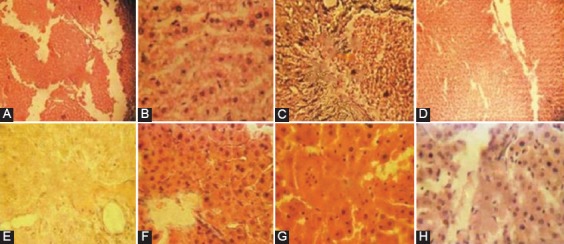
Figure 1.

Photomicrographs of liver tissue (section stained with H&E, ×400) (A) CTRL: No visible lesions, (B) QU: Diffuse hydropic degeneration of the hepatocytes with very prominent sinusoids, (C) CH: Marked portal congestion and diffuse vacuolar degeneration of the hepatocytes, (D) CHQU: Mild Kupffer cell proliferation with moderate hepatic vacuolar degeneration, associated with moderate periportal cellular infiltration by mononuclear cells, (E) CHPG(1): Mild congestion of the portal vessels, with very mild periportal, hydropic hepatic degeneration, (F) CHPG(2): Mild portal congestion and mononuclear cellular infiltration, mild periportal and hydropic degeneration, (G) CHSI(1): Marked portal congestion and cellular infiltration by mononuclear cells, (H) CHSI(2): Portal canal congestion moderately infiltrated by mononuclear cells.
