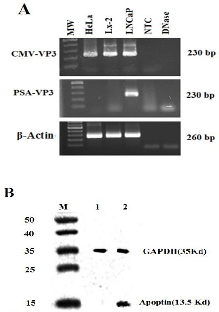
Figure 3.

Specific expression of apoptin in LNCaP cells transfected with PSES-pAdenoVator-PSA-Apoptin-IRES-GFP. A: RT-PCR performed with apoptin-specific primers showed that apoptin was expressed in LNCaP cells that transfected with PSES-pAdenoVator-PSA-Apoptin-IRES-GFP, whereas the non-prostatic cell lines (LX-2 and HeLa cell lines) were negative for apoptin expression, indicating by absence of 230 bp band. β-actin expression was used as a reference gene in each sample. B: Western blot showed the presence of apoptin (13.5 KDa) in LNCaP cells transfected with PSES-pAdenoVator-PSA-Apoptin-IRES-GFP (Lane 2) but not in non-transfected cells (Lane 1). GAPDH expression was used as an internal control in each sample. M indicates the protein molecular weight marker (15-50 KDa)
