Figure 6.
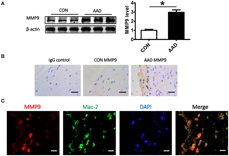
The correlation between FKBP11 and MMP9 expression in infiltrated macrophages under AAD. (A) Representative immune-blots (upper panel) and bar graphs (lower panel) showing MMP9 expression levels in whole aorta tissue lysates from AAD patients and control group. (B) Exemplary immune-histochemical staining of aortic paraffin embedded tissue sections from healthy donors and AAD patients; left, IgG control; middle, healthy donor; right, AAD patient; MMP9 antigen detected by HRP-DAB reaction, brown; hematoxylin counterstain, blue; scale bar: 100 μm. (C) Double immunofluorescent staining showed co-localization of MMP9 with macrophage marker Mac-2. It showed that the expression of MMP9 in infiltrated macrophages increased in AAD group. MMP9 staining (red), nuclear stain DAPI (blue), macrophage marker (Mac-2) staining (green); scale bar: 100 μm. CON, control healthy donor, open bars; AAD, patient with acute aortic dissection, black bars. Mean ± SEM of 6 subjects per group (A–C); *P < 0.05 (A, student t-test).

