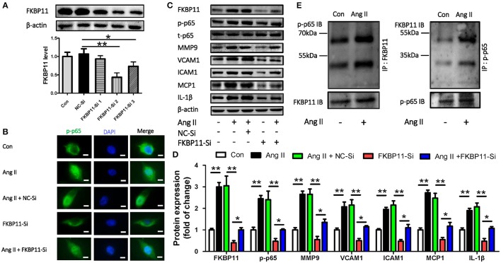
Figure 7.
The pro-inflammatory function of FKBP11 and activation of NF-kB p65 subunit in endothelial cell. (A) Representative immunoblots (upper panel) and bar graph (lower panel) showed FKBP11 specific siRNA FKBP11-Si2 and FKBP11-Si3 but not FKBP11-Si1 can effectively suppress the protein expression as compared to the scrambled control NC-Si in EA.hy926 cells. (B–D) EA.hy926 cells were incubated with Ang II after transfected with FKBP11-Si2 or scrambled control NC-Si. (B) Cytofluorescent picture showed that Ang II promoted the nuclear localization of p-p65, while FKBP11 siRNA treatment could blunt it. Together, representative immunoblots (C) and bar graph (D) showing FKBP11-siRNA treatment could effectively suppress the phosphorylation and nuclear translocation of p65 and subsequently the expression of pro-inflammatory cytokines MCP1, VCAM1, ICAM1, and IL1-β. p-p65 staining (green); nuclear stain DAPI (blue); scale bar: 100 μm. (E) The interaction of FKBP11 and p-p65 was enhanced following Ang II treatment in EA.hy926 cells. Detection of p-p65 by immunoblotting following immunoprecipitation of FKBP11 from whole cell lysates of control (Con) cells or of cells after Ang II treatment (Ang II), left panel or vice versa, right panel; representative images of precipitated protein immunoblots (top) and respective input immunoblots (bottom). IgG controls for immunoprecipitation experiments were attached to the Supplementary Figure 3. CON: Control cells without treatment; Ang II: 1.0 × 10⁻⁶ mol/L Angiotensin II treated cells; NC-Si, Scrambled SiRNA for FKBP11; FKBP11-Si, FKBP11 knockdown SiRNA. Mean value ± SEM of at least three independent experiments (A,D); *P < 0.05; **P < 0.01 (ANOVA); Representative immunoblots of at least three independent experiments; (A,C,E); IB, immunoblot; IP, immunoprecipitation.

