Figure 8.
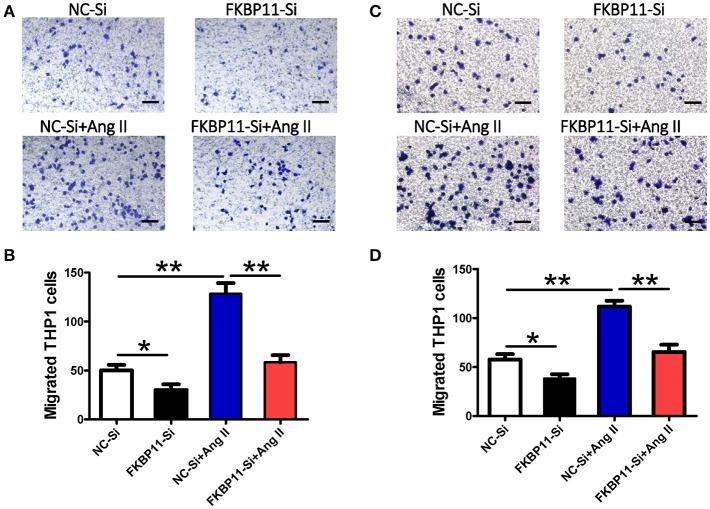
FKBP11 and monocyte transmigration through an endothelial cell monolayer. (A,B) EA.hy926 monolayer in the lower compartment was incubated without or with Ang II after transfected with FKBP11-Si RNA or NC–Si RNA for 24 h. Then 5 × 10⁵ THP-1 cells were seeded into each upper compartment containing 100 μL serum-free RPMI medium. Images depicting crystal violet stained THP-1 cells which migrated across the membrane of transwell inserts (pore size 8 μm) to the lower side after incubation with conditioned media from the lower compartment for 12 h. Representative pictures are shown. (A) Migrated cells per high power field were quantified and the data were showed in bar graph (B). (C,D) The same as in (A,B), instead of EA.hy926 cell monolayer, HUVECs were applied. NC-Si: Scrambled SiRNA for FKBP11 treated endothelial cells; FKBP11-Si: FKBP11 knockdown SiRNA treated endothelial cells; NC-Si+Ang II: Scrambled SiRNA for FKBP11 and 1.0 × 10⁻⁶ mol/L Angiotensin II treated endothelial cells; FKBP11-Si+AngII: FKBP11 knockdown SiRNA and 1.0 × 10⁻⁶ mol/L Angiotensin II treated endothelial cells. Mean value ± SEM of at least three independent experiments (B,D); *P < 0.05; **P < 0.01 (ANOVA); Scale bar: 200 μm.

