Fig. 3.

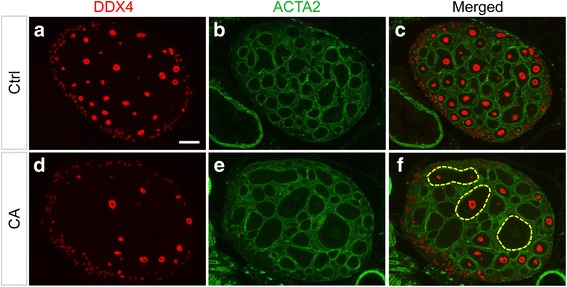
Immunofluorescence analysis of follicular defects in TGFBR1-CAG9Cre mice. a-f Double immunofluorescence of DDX4 (red) and ACTA2 (green) using PD12 control (a-c) and TGFBR1-CAG9Cre (d-f) mice. Three independent samples per group were analyzed using immunohistochemistry and/or immunofluorescence. Note the presence of large follicles or abnormal follicle-like structures (dotted yellow lines) in the ovaries of TGFBR1-CAG9Cre mice in comparison with age-matched controls. Scale bar is representatively depicted in (a), and equals 100 μm (a-f)
