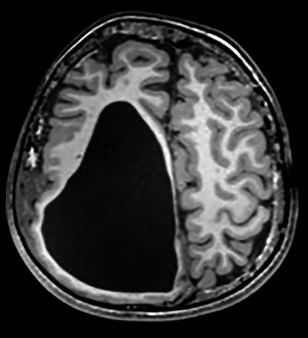
Figure 3.

Axial T1A cranial MRI image of asymmetric cystic dilatation of the right lateral ventricle occipital horn, deletion of gyri and sulci irregularity with cortical thinning and diffuse white matter volume decrease, intracranial lipoma under the skin.
