
Figure 4.
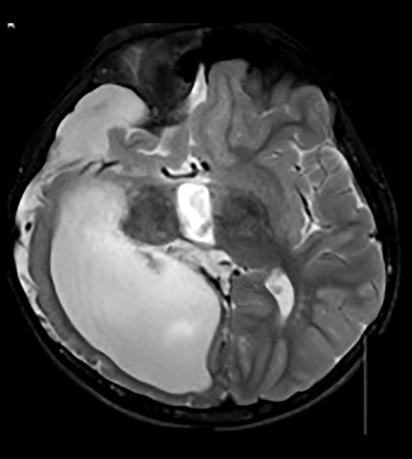
Axial T2A cranial MRI image of asymmetric cystic dilatation of the right lateral ventricle occipital horn, deletion of temporo-occipital gyri and sulci irregularity with cortical thinning and diffuse white matter volume decrease and subdural effusion.