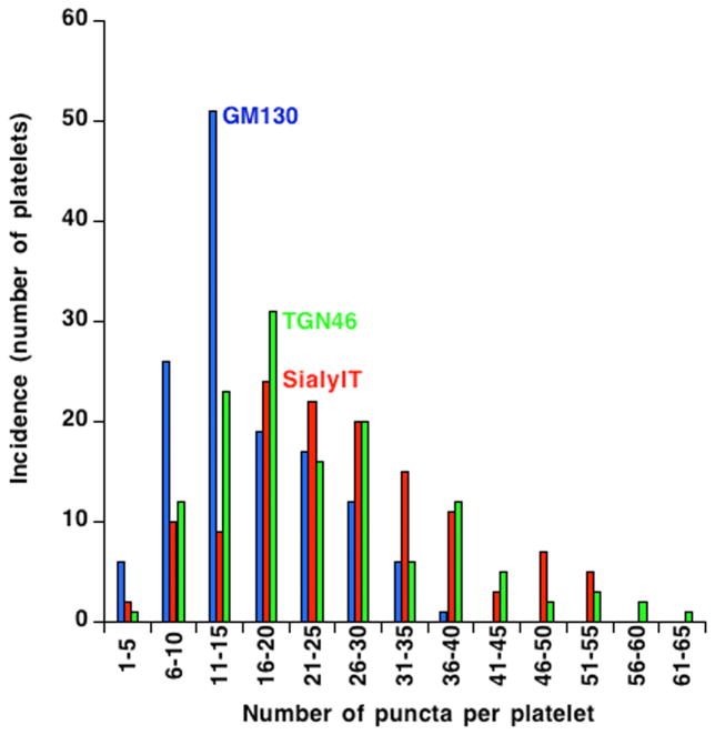
Figure 5. The incidence of Golgi-protein-positive puncta in resting human platelets suggest that circulating platelets behave as a single population with respect to Golgi organization.
The incidence of Golgi-protein-positive puncta were manually calculated for 3 Golgi markers GM130 (blue), SialylT (red) and TGN46 (Green) using deconvolved, widefield images of 135 platelets. The frequency distribution of puncta per platelet is shown as histograms using KaleidaGraph software version 4.5.2. The histogram plots of each of the three markers are smooth and fairly similar suggesting that circulating platelets behave as a single population.

