
Figure 3.
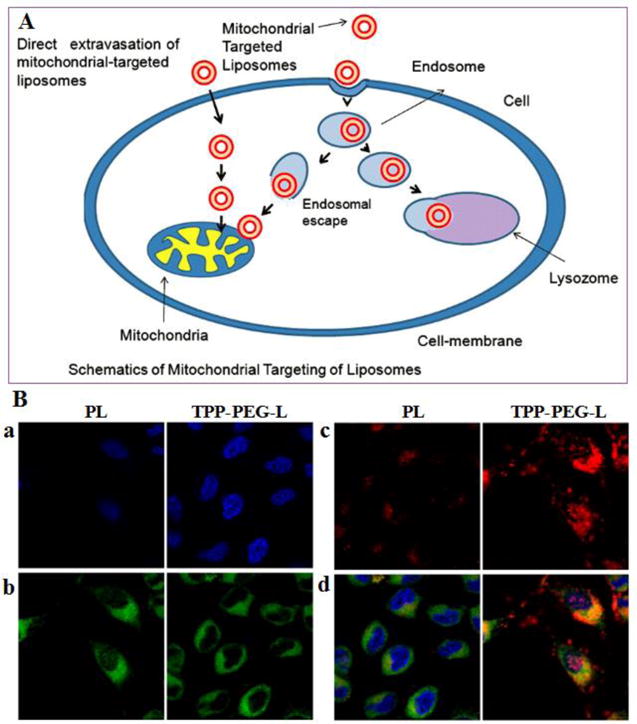
(A) Schematic illustration of mitochondrial targeting of TPP-PEG-L. (B) Mitochondrial colocalization of fluorescently-labeled TPP-PEG-L compared to PL by confocal laser scanning microscopy. HeLa cells were incubated with Rh-PE-labeled TPP-PEG-L and PL for 18 h and then stained with MTG and Hoechst 33342. Yellow spots in the merged pictures denote the co-localization of the liposomes within mitochondrial compartments. a. Images of nuclei stained with Hoechst; b. Mitochondria staining with MTG; c. Uptake of Rh-labeled liposomes; d. Merged picture of all. (Reproduced from ref. [42] with permission)