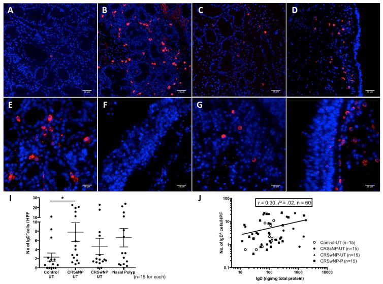
Figure 2.
Immunofluorescence analysis of IgD+ cells in nasal tissue by using phycoerythrin-conjugated anti-IgD (red). Representative images of IgD+ cells in UT from A, control, B, CRSsNP, C, CRSwNP, and D, NP from CRSwNP patients (magnification 200x). E, IgD+ cells at the peri-glandular and F, subepithelial area in CRSsNP UT and G, peri-glandular and H, subepithelial area in NP of CRSwNP (magnification 400x). Nuclei were counterstained with 49,6-diamidino-2-phenylindole (blue). I, IgD+ cells in nasal tissue were counted semiquantitatively. J, Correlation between the numbers of IgD+ cells from immunofluorescence assay and IgD protein concentrations in matched nasal tissue. *P < .05. HPF, High-power field.

