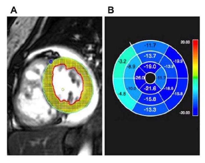
Fig. 1.

A) Feature-tracking strain analysis software using cardiac magnetic resonance, with representative short axis Steady State Free Precession (SSFP) image demonstrating myocardial tracking
B) Software output demonstrating abnormal peak circumferential strain (light blue/green indicate abnormal strain as indicated by negative numbers closer to zero) in the ventricular septum in a patient with pulmonary hypertension
