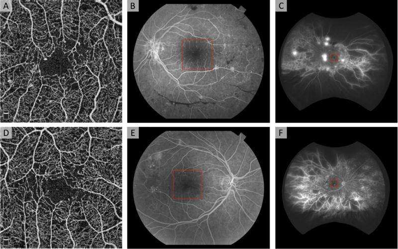
Figure 3. Field of View Limitations in OCTA Shown in Two Eyes with Proliferative Diabetic Retinopathy.

(A, D) En face 3 × 3 mm2 OCTA image of the superficial capillary plexus reveals mild diabetic changes including enlarged foveal avascular zone, areas of non-perfusion, and microaneurysms. (B, E) Fluorescein angiography obtained with fundus camera (50° field of view) reveals hemorrhages and neovascularization (NV) in B, and non-perfusion and NV in E. (C, F) Fluorescein angiography obtained with Optos wide-field imaging (200° field of view) reveals extensive peripheral non-perfusion and NV.
