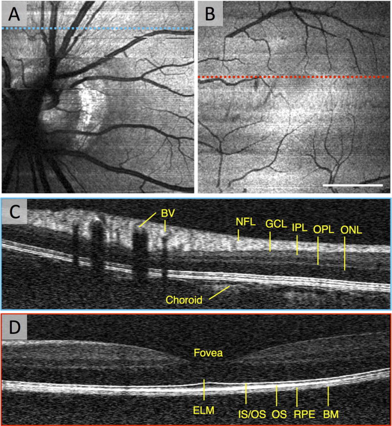
Figure 8. Structural Imaging of the Human Macula with vis-OCT.

(A) An en face vis-OCT image of the optic nerve head in a healthy 23 year old volunteer. (B) An en face vis-OCT image of the macula in the same volunteer. (C) The vis-OCT B-scan of some major vessels superior to the optic nerve head corresponding to the blue dashed line in (A). (D) The vis-OCT B-scan of the fovea corresponding to the red dashed line in (B). BV: blood vessel. NFL: nerve fiber layer. GCL: ganglion cell layer. IPL: inner plexiform layer. OPL: outer plexiform layer. ONL: outer nuclear layer. ELM: external limiting membrane. IS/OS: inner segment/outer segment junction. OS: outer segments. RPE: retina pigment epithelium. BM: Bruch’s membrane. Scale bar: 2 degrees.
