Figure 1.
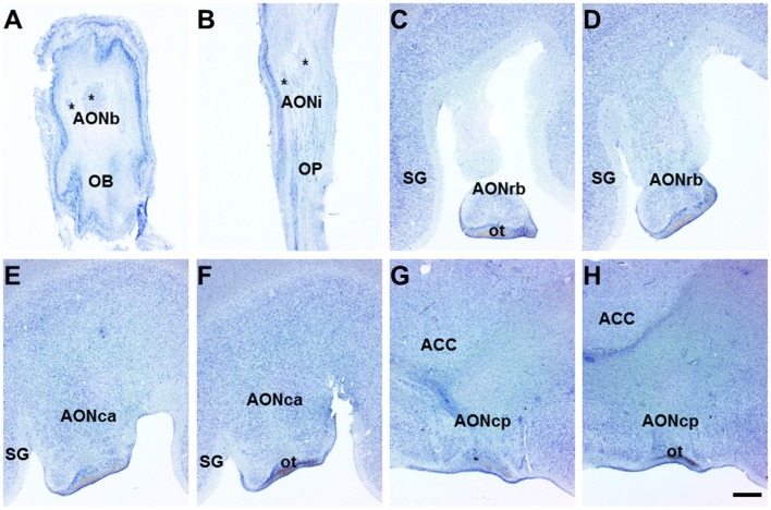
Horizontal (olfactory bulb (OB) and olfactory peduncle (OP)) and coronal (frontal lobe) sections of the human brain. Nissl-stained horizontal sections of the human OB and OP show bulbar (A) and intrapeduncular (B) anterior olfactory nucleus (AON) showing diverse components (asterisks). In coronal sections of the frontal lobe, the retrobulbar (C,D), cortical anterior (E,F) and cortical posterior (G,H) AON are distinguishable. Scale bar, 1300 μm. For abbreviations, see list.

