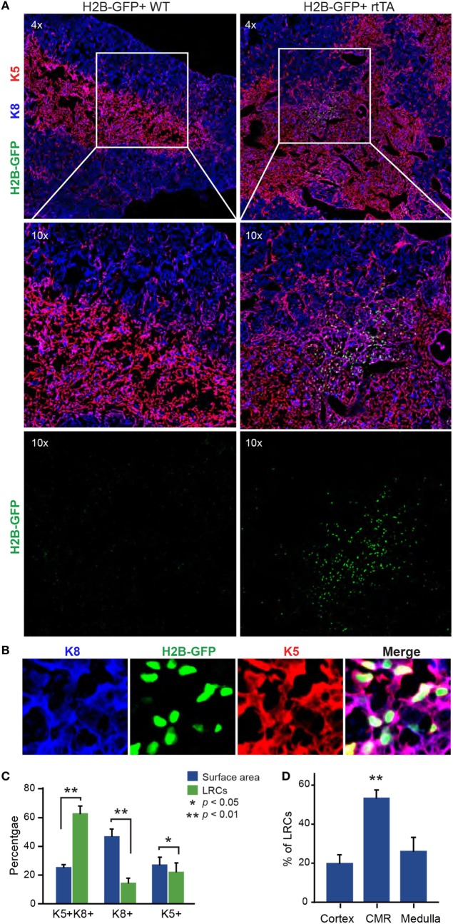
Figure 1.
Most label-retaining cells (LRCs) are K5+K8+ and localized near the cortico-medullary junction. (A) Representative images of thymic slices after 16 weeks of chase in control (H2B-GFP+ WT, left panels) and test mice (H2B-GFP+ rtTA, right panels). (B) Representative images of K5+K8+ GFPhi cells. Cytokeratin K5 is shown in red, K8 in blue, and histone 2B-GFP fusion protein (H2B-GFP) in green. (C) Quantification of LRCs expressing K5, K8, or both cytokeratins. The number of LRCs per thymic lobe (green) from each TEC subset is compared with the surface area (blue) covered by this subset. (D) Percentage of LRCs found in different regions of the thymus. The proportions of LRCs are significantly higher in the cortico-medullary region (CMR) than the cortex or the medulla (n = 4).

