Figure 3.
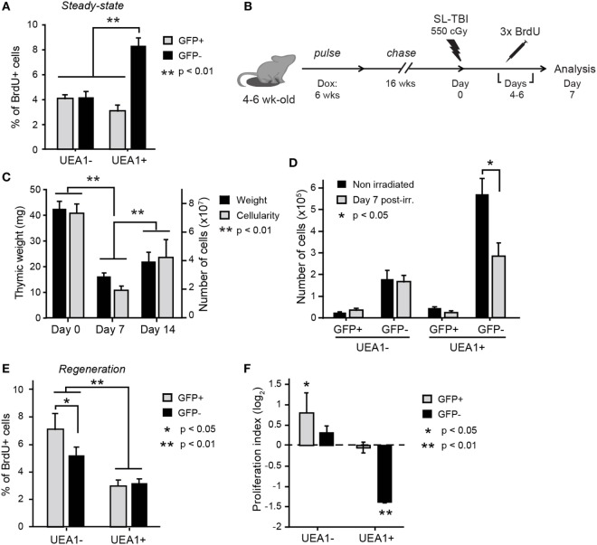
UEA1– label-retaining cells (LRCs) participate to thymic regeneration following SL-TBI. (A) Percentage of BrdU+ thymic epithelial cells (TECs) under steady-state conditions. Statistical differences were calculated using paired Student’s t-test (n = 10). (B) Schematic representation of the thymic injury assay. (C) Thymic weight (black) and cellularity (gray) at different times before and after irradiation (n = 4–11 per group). (D) Number of cells in each TEC subset in non-irradiated controls (day 0, black) and during regeneration following SL-TBI (day 7, gray, n = 5). Percentage of BrdU+ TECs (E) and proliferation index (F) for each subpopulation of TECs during thymic regeneration. The proliferation index is calculated using the following equation: proportion of BrdU+ cells during regeneration (day 7)/proportion of BrdU+ cells before SL-TBI (day 0). Cells derived from LRCs (labeled GFP+) or NonLRCs (labeled GFP–) are represented in gray and black, respectively (n = 9–10 per group). Statistical differences for panels (C,D,F) were calculated by cell population, comparing day 0 non-irradiated to day 7 postirradiation or day 14 to day 7 postirradiation. Data are represented as mean + SEM.

