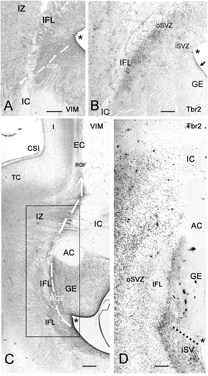
FIGURE 8.

Photographic reconstructions of the PSB in coronal sections at 21 GW. Vimentin (A,C) shows the distribution and orientation of radial glia cells and fibers in the frontal (A) and temporal (C) periventricular zones and the RGF route into the Insula (white arrows). Tbr2 (B,D) marks the PSB (arrow in B), which at this age is less well defined than at earlier fetal stages, and in the temporal lobe does not extend medially beyond the striato-cortical sulcus (asterisks in all panels; in D, the PSB is indicated by a dotted line). Tbr2+ and vimentin+ progenitor cells cross the periventricular fiber layers (Inner fibrous layer, IFL) and extend far into the oSVZ and even the intermediate zone (IZ). Note the complexity of fiber tracts in the temporal lobe, due to the presence of the anterior commissure (AC). The large black dots in the inner (i) SVZ in D are stained blood vessels. Bars: in A: 240 μm, in B: 140 μm, in C: 250 μm, in D: 190 μm.
