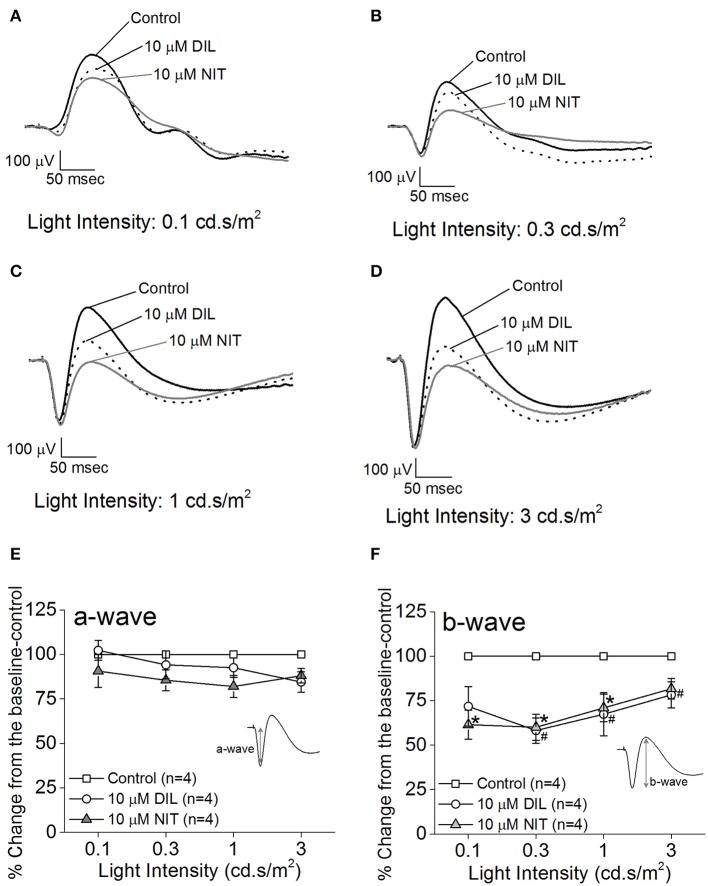
Figure 2.
Differential contributions of LTCCs to retinal light responses as measured by ex vivo ERGs. Mice were dark adapted for at least 3 h, and the retinas were excised and placed in an ex vivo ERG recording chamber. The ERG responses were recorded under 4 different light intensities: 0.1, 0.3, 1, and 3 cd·s/m2. The ERG recordings were performed with normal perfusion buffer (Control), followed by perfusion with 10 μM DIL to inhibit Cav1.2, and subsequently perfused with 10 μM nitrendipine (NIT) to block both Cav1.2 and Cav1.3. (A–D) Representative ERG waveforms recorded in different solutions (control, 10 μM DIL, and 10 μM NIT) are shown, which were recorded under light intensities of 0.1, 0.3, 1, and 3 cd·s/m2, respectively. (E) Perfusion with 10 μM DIL or 10 μM NIT for 10 min did not have significant effect on the ERG a-wave amplitudes. (F) Perfusion with 10 μM DIL or 10 μM NIT for 10 min decreased ERG b-wave amplitudes. The asterisk (*) indicates a statistically significant difference between the control and the 10 μM NIT group; “#” indicates that the 10 μM DIL group is statistically different from the control. *, #p < 0.05.

