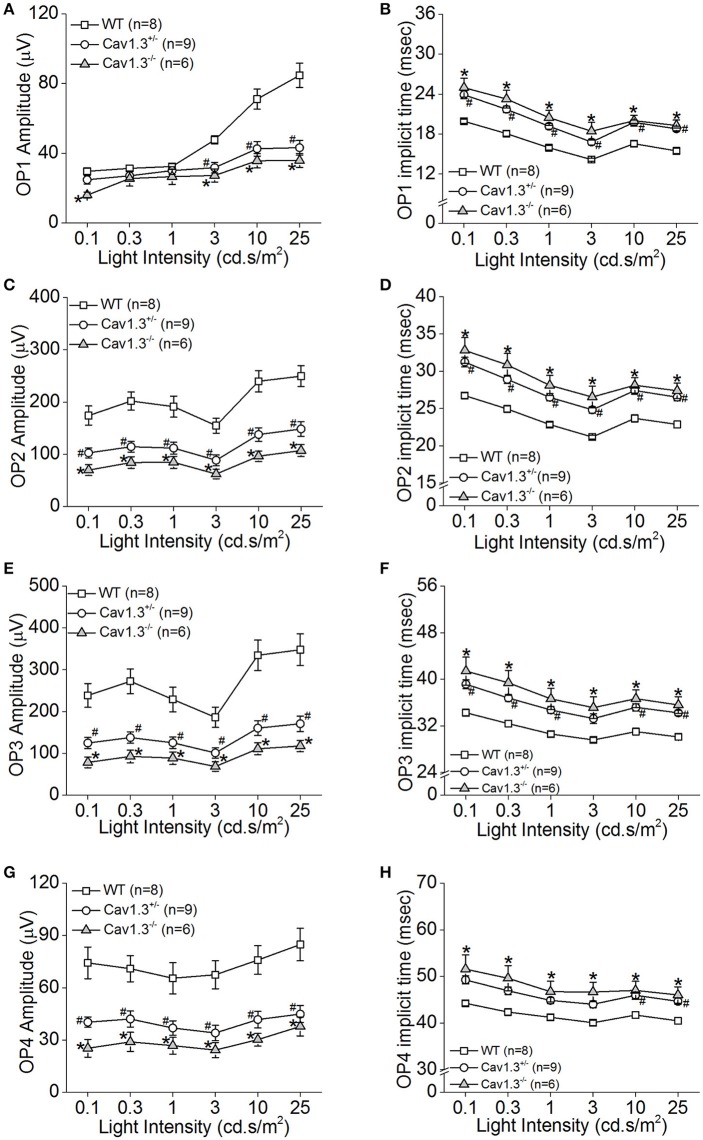
Figure 5.
The inner retinal light responses are decreased in Cav1.3+/− and Cav1.3−/− mutant mice. The oscillatory potential (OP) amplitudes are decreased and implicit times delayed in both Cav1.3+/− and Cav1.3−/− mice compared to the wild type (WT). The OPs were analyzed after the scotopic ERG responses were filtered between 100 and 300 Hz band-pass using the ERGView4.4 software (Ocuscience). (A,B) The average OP1 amplitudes (A) are decreased, but the implicit time (B) is increased in Cav1.3+/− and Cav1.3−/− mice compared to the WT. (C,D) The average OP2 amplitudes (C) are decreased, but the implicit time (D) is increased in Cav1.3+/− and Cav1.3−/− mice compared to the WT. (E,F) The average OP3 amplitudes (E) are decreased, but the implicit time (F) is increased in Cav1.3+/− and Cav1.3−/− mice compared to the WT. (G,H) The average OP4 amplitudes (G) are decreased, but the implicit time (H) is increased in Cav1.3+/− and Cav1.3−/− mice compared to the WT. The asterisk (*) indicates a statistically significant difference between the WT and Cav1.3−/− mice; “#” indicates a statistically significant difference between the WT and Cav1.3+/− mice. *, #p < 0.05.

