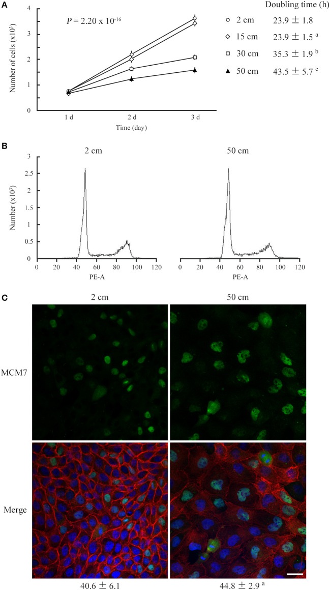
Figure 1.
Low static pressure suppresses MDCK cell number increase. (A) MDCK cells were cultured on a semipermeable membrane in 2-, 15-, 30-, or 50-cm-high culture medium for 3 days. Cell numbers were counted every day, and the means were plotted with error bars indicating standard deviations (n = 3 per group for each experiment). The time-course plots of cell numbers were analyzed by ANOVA; the P-value is shown. Cell doubling time (mean ± standard deviation, hour) was calculated from five independent experiments. a,b,cP = 0.985, < 0.001, and < 0.001, respectively, by Student's t-test when compared with 2-cm-high-medium cultures. (B) After 3 days of culture in 2- or 50-cm-high medium, MDCK cells were labeled with propidium iodide and analyzed by flow cytometry. Representative results are shown. (C) MDCK cells were cultured in 2- or 50-cm-high medium for 3 days, then were triple-stained with MCM7 immunofluorescence (green; upper), phalloidin labeling (red), and DAPI nuclear staining (blue). The three images are merged (lower). MCM7 positivities (mean ± standard deviation, %; n = 3 per group) are shown below the photo panels. aP = 0.208, by Student's t-test when compared with 2-cm-high-medium cultures. Scale bar = 20 μm.

