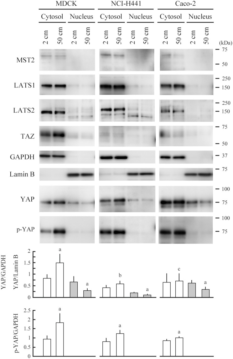
Figure 3.
Expression analyses of Hippo pathway molecules. MDCK, NCI-H441, and Caco-2 cells were cultured on a semipermeable membrane in 2- or 50-cm-high medium for 3 days. Cytosolic and nuclear proteins were separately extracted from the cells, and were blotted with the antibodies indicated. Glyceraldehyde 3-phosphate dehydrogenase (GAPDH) and lamin B were used as cytoplasmic and nuclear markers, respectively. Intensities of the immunoreactive bands for YAP, serine-127 phosphorylated YAP (p-YAP), GAPDH, and lamin B were measured densitometrically. The mean ratios of cytosolic YAP and p-YAP to GAPDH (white bar) and nuclear YAP to lamin B (gray bar) and their standard deviations were calculated from the data obtained in three independent experiments. aP < 0.05, and b,cP = 0.213 and 0.868, respectively, by Student's t-test when compared with 2-cm-high-medium cultures.

