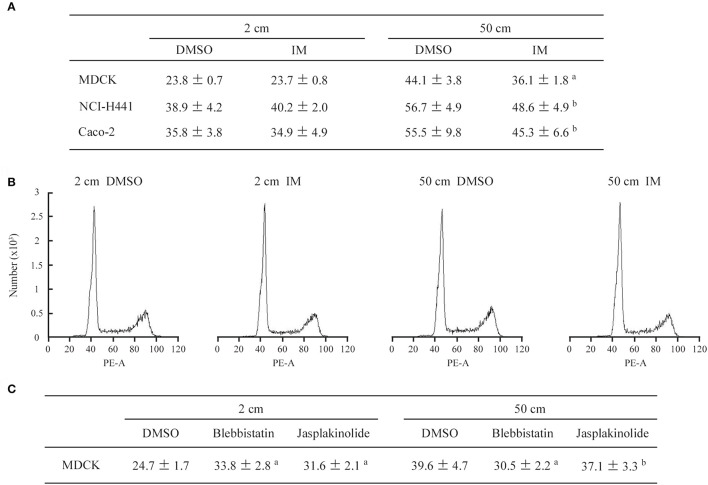
Figure 5.
Cell growth and cell cycle analyses in the presence of pharmacological agents. (A) MDCK, NCI-H441, and Caco-2 cells were cultured in 2- or 50-cm-high medium containing either irsogladine maleate (IM; 100 nM) or DMSO (10,000× dilution), and their doubling times were calculated (n = 3 per group for each experiment). The mean and standard deviation (hour) of five independent experiments are shown. a,bP ≤ 0.01 and 0.05, respectively, by Student's t-test when compared with the 50 cm DMSO group. (B) After 3 days of culture in 2- or 50-cm-high medium, MDCK cells were labeled with propidium iodide and analyzed by flow cytometry. Representative results are shown. (C) MDCK cells were cultured in 2- or 50-cm-high medium containing blebbistatin (20 μM), jasplakinolide (20 nM) or DMSO (7,500 × dilution), and their doubling times were calculated (n = 3 per group for each experiment). The mean and standard deviation (hour) of five independent experiments are shown. a,bP ≤ 0.01 and P = 0.371, respectively, by Student's t-test when compared with the 50 cm DMSO group.

